June 11, 2024
Share
Scientists continue to make significant strides in cancer research with the help of cutting-edge approaches like 3D genomics. By analyzing the three-dimensional organization of the genome, researchers can uncover how genetic and epigenetic changes drive cancer development and progression.
This year, we received an impressive variety of project proposals for the 2024 Cancer Research Grant, showcasing the innovative thinking of researchers in the field as well as the expanding applications for 3D genomics in cancer research. After a thorough review, the Arima grant committee is delighted to announce the 5 recipients of this year’s cancer research grants! Read on to learn about the groundbreaking projects our grant recipients will be pursuing.
Detecting Structural Variants in Adenoid Cystic Carcinoma
Grant Recipient: Elisabet Pujadas, MD, PhD

Elisabet Pujadas, MD, PhD of Icahn School of Medicine at Mount Sinai and Memorial Sloan Kettering Cancer Center
Institution: Icahn School of Medicine at Mount Sinai and Memorial Sloan Kettering Cancer Center
Project Title: Structural variant detection in adenoid cystic carcinoma: the old and the new
Project Overview: Adenoid cystic carcinoma (AdCC) is a highly infiltrative malignancy most commonly affecting the head and neck that harbors pathognomonic t(6;9) MYB::NFIB and t(8;9) MYBL1::NFIB fusions. Detection of these fusions using FISH or RNA sequencing methods can be a key aspect in rendering a pathologic diagnosis, especially in tumors with challenging morphology. Unfortunately, up to a third of cases lack such defining fusions and the high variability of breakpoints for known fusions makes assay design difficult.
For this project, the goal of Elisabet Pujadas’ lab is to demonstrate the clinical utility of Hi-C for detecting structural variants in archival FFPE diagnostic samples by showing concordance with previously detected AdCC fusions as well as undertaking an unbiased, genome-wide search of possible novel structural variants in AdCC where canonical fusions were not detectable using conventional techniques. This approach could pave the way for similar translational studies in other fusion-driven malignancies across organ-sites.
“Hi-C has a strong track record in the field of epigenomics and is reaching a point in its development where we can start thinking about its translational use in archival clinical samples in a scalable fashion. I am very interested in utilizing Hi-C as a discovery tool in the space of structural variants, where unbiased, genome-wide tools are currently lacking.” – Elisabet Pujadas, MD, PhD
DNA Damage Response and 3D Genome Organization in SCLC
Grant Recipient: Carminia Della Corte, PhD

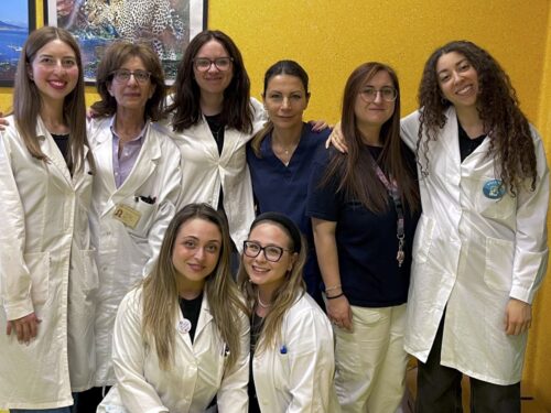
Precision Medicine Department, University of Campania Luigi Vanvitelli
Institution: University of Campania Luigi Vanvitelli
Project Title:The crosstalk between DNA damage and 3D genome organisation role in mediating response to chemo-immunotherapy in SCLC
Project Overview: Many recent studies have reported a high genetic expression of different DNA damage response (DDR) proteins in small cell lung cancer (SCLC). The 3D architecture of the genome, DNA repair factors and cytoskeleton work together in promoting DDR signaling and damaged DNA mobility and repair. The Precision Medicine Department at the University of Campania Luigi Vanvitelli aims to assess the 3D genomic data to model the clinical effectiveness of combinations of DDR inhibitors (DDRi) and anti-PD-L1 in SCLC models. They will test DDRi in SCLC patient-derived circulating tumor cells (CTCs) and immune cells obtained from treatment-naive and chemo-immunotherapy relapsed SCLC patients in order to predict the role of DDRi in the clinical setting of second-line treatment. Using Hi-C, the team will investigate the organizational structure of chromatin in the crosstalk between DDR and 3D genome organization after treatment with DDRi + anti-PD-L1, dissecting its effect in orchestrating post-treatment changes in innate pathway activation.
“Thanks to the Arima Genomics Cancer Research Grant, Hi-C will enable the study of how DNA damage response and repair affect 3D genome folding. Using selective DNA damage response (DDR) inhibitors, we will assess changes in 3D genome structural integrity in circulating tumour cells and immune cells derived from SCLC patients. Hi-C/3D genomics will help to uncover potential changes in genome-wide 3D organisation as a possible mechanism of response or resistance to therapy.” – Dr. Caterina De Rosa
Epigenetic Changes During Progression of IDH-mutant Astrocytomas
Grant Recipient: Abigail Suwala, MD

Abigail Suwala, MD, University Hospital Heidelberg and DKFZ
Institution: University Hospital Heidelberg and DKFZ
Project Title: Deciphering changes in chromatin conformation during the progression of IDH-mutant gliomas
Project Overview: The Suwala Lab is studying malignant brain tumors called IDH-mutant astrocytomas that mostly affect younger adults. These tumors often reoccur after initial treatment, evolving into a more malignant state with dismal prognosis. During progression, tremendous changes in DNA methylation are observered, indicating that epigenetic transformations play a role in progression. The Suwala Lab is highly interested in analyzing how chromatin structure changes over time and treatment, and how it can be linked to DNA methylation changes, shedding light into molecular mechanisms for future targeted treatments.
“We are excited to use the Arima-HiC FFPE Kit in our research. As human biopsy samples are mostly stored as FFPE tissue blocks, this gives us the chance to study chromatin structure in “real” brain tumors rather than models.” – Abigail Suwala, MD, PhD
Analyzing 3D Chromatin Architecture in Bioprinted Tumor Models
Grant Recipient: Shreyas Gaikwad, PhD Student

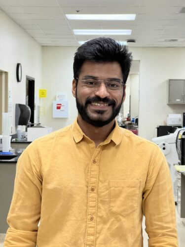
Shreyas Gaikwad, Texas Tech University Health Science Center.
Institution: Texas Tech University Health Science Center
Project Title: Bioprinted tumor models: Comparing the omics between flat biology and 3D world of cancer cells
Project Overview: This project aims to analyze 3D chromatin architecture changes in cells cultured within 3D bioprinted tumor models compared to conventional 2D culture systems using Hi-C technology. Hi-C, a powerful tool for mapping chromatin interactions, will enable a comprehensive understanding of the spatial organization of the genome in different culture conditions. By comparing chromatin interaction maps from cells in 3D bioprinted environments to those in 2D cultures, Shreyas Gaikwad of the Srivastava Lab aims to elucidate how the 3D cellular microenvironment influences genomic organization and gene regulation. This analysis is crucial for understanding tumor biology as the 3D tumor models better mimic the in vivo conditions of solid tumors, providing more physiologically relevant insights. The outcomes could reveal novel regulatory mechanisms driving tumor progression and identify potential therapeutic targets by highlighting differences in chromatin structure associated with the 3D culture environment.
“I am excited about 3D genomics and Hi-C in my project, as this offers unprecedented insights into the spatial organization of the genome. As we delve more into 3D models as tools for drug discovery, it is important to understand how close are these models to the real in vivo setting.” – Shreyas Gaikwad, PhD Student.
Exploring the Role of Steroid Receptors in Breast Cancer
Grant Recipient: Wendy Effah, PhD Candidate

Wendy Effah, University of Tennessee Health Science Center
Institution: University of Tennessee Health Science Center
Project Title: Context-dependent role of glucocorticoids in breast cancer
Project Overview: Glucocorticoids bind to the transcription factor glucocorticoid receptor (GR) and regulate genes in response to cellular metabolism, inflammation, and stress. Although GR has been extensively evaluated in inflammation and metabolic diseases, its role as a therapeutic target in cancer has not been extensively studied. Results with the androgen receptor (AR), another member of the steroid receptor family, demonstrated that the AR is an oncogene in estrogen receptor (ER)-negative breast cancer, but a tumor suppressor in ER-positive breast cancer. Wendy Effah of the Narayanan Lab expected a comparable mechanism with GR in different subtypes of breast cancer subtypes. In a previous study, the researcher evaluated the role of GR in various breast cancer subtypes and discerned the mechanism of action.
The objective of the study is to develop alternate approaches utilizing steroid receptors to treat therapy-resistant breast cancer. Preliminary results led to the central hypothesis that steroid receptor ligands have tumor growth inhibitory properties and appropriate utilization will render a new therapeutic target that will result in sustained tumor growth inhibition. 3D genomics will be insightful in explaining why the receptors act differently in different subtypes of breast cancer.
“The area of 3D genomics excites me for its in-depth visualization of genome architecture in processes like DNA replication and transcription factor regulation.” – Wendy Effah, PhD Student
Congratulations again to these remarkable researchers! Stay tuned for updates on the exciting progress of our 2024 Cancer Research Grant recipients, and learn how 3D genomics can play a pivotal role in your cancer research.


